

- Home
- Units
- Light, nanomaterials, nanotechnologies (L2n - CNRS-UMR 7076)
- Research topics
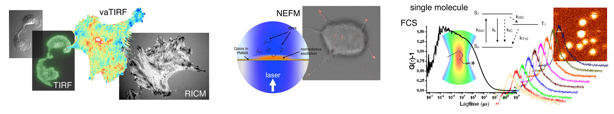
- Biophotonics and nanosensors
vaTIRF nanoscopy
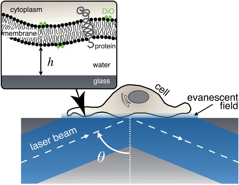
Variable-angle Total Internal Reflection Fluorescence nanoscopy is a powerful technique to observe the cell cortex (plasma membrane, actin cytoskeleton, vesicle trafficking…). We use this nanoimaging method to quantify the adhesion of living cells and to study the interaction between the cell membrane and its environment. Since several decades, TIRF microscopy is commonly used to observe cell dynamics, because it avoids phototoxicity by limiting cell exposure to laser irradiation. Only the cell cortex in contact with the substrate, within a few hundred nanometers thick, is excited by the evanescent field in TIRF imaging.
Research
NEF nanoscopy
vaTIRF nanoscopy
nanoFCS
RICM
Cell patterning
Publications
Collaborations
vaTIRF is an old technique introduced in the mid-80s and quickly forgotten due to the high complexity of the first experimental setup which used a prism to create the evanescent wave. We recently proposed an improved straightforward version of vaTIRF adapted to modern TIRF setup. We have demonstrated that a series of 10 TIRF images recorded at different incident angles θ are useful to reconstruct the cell membrane topography of motile cells with a nanometric axial precision.
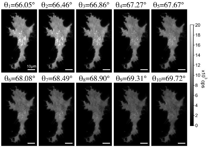
vaTIRF images recording of the same U87MG cell in adhesion on fibronectin (the plasma membrane is labeled with DiO).
The extreme axial precision achievable with this method, 20-30 nm, is remarkable and well adapted to localize precisely the height h of the plasma membrane and thus probe cell membrane/substrate interactions. But, vaTIRF also permits to measure the effective refractive index, denoted neff, of the cell cortex within the evanescent field. neff depends on different parameters, and it will notably increase together with the cytoplasmic refractive index. The interactions between ligands and receptors (for example integrins and fibronectin binding) affect significantly the height h of the membrane, which most closely approaches the substrate on focal adhesion zones. Moreover, the functional unit of focal contacts includes a lot of intracellular proteins. This gives rise to a significant increase of the local cytoplasmic refractive index. Hence, neff will be higher on focal adhesion zones due to the presence of various proteins such as actin, FAK, vinculin, paxillin, talin... Finally, focal adhesion and more generally adhesion plaque should appear where the plasma membrane closely approaches the substrate and the effective refractive index is high. An example of focal adhesion localization is given in the figure below.
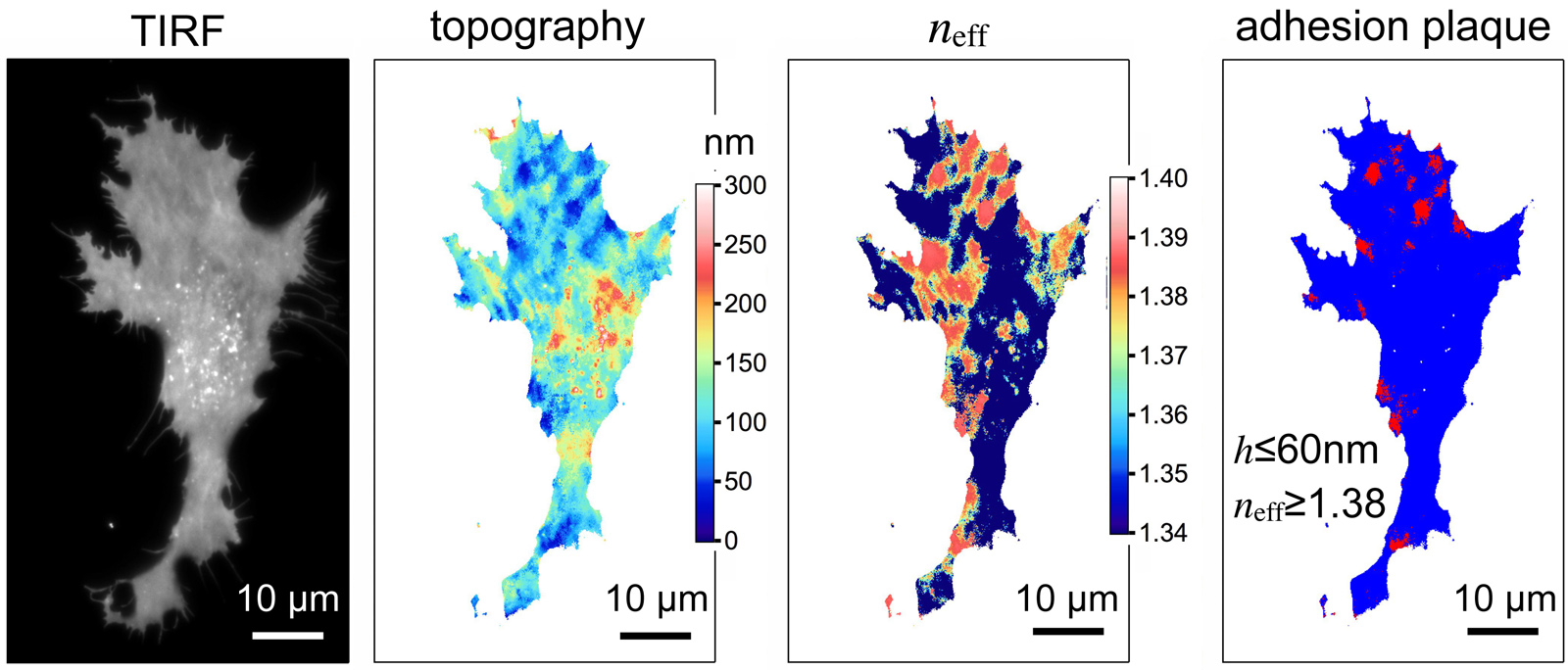
Example of vaTIRFM reconstruction on U87MG cell in adhesion on fibronectin. From the left to the right: TIRF image, the corresponding cell membrane topography, neff image and 2D localization of the focal adhesion zones which appear in red color.