
- Home
- Units
- Light, nanomaterials, nanotechnologies (L2n - CNRS-UMR 7076)
- Research topics
- Light-Matter interactions at the nanoscale
Chiral plasmonics
Principal investigator: Davy Gérard
Summary
An object is chiral if it cannot be superimposed to its mirror image. Peculiar properties, known as optical activity, appears when light (notably polarized light) interacts with a chiral object. Although ubiquitous in the natural world, "natural" chirality generally leads to weak chiroptical interactions. In this research project, we design artificial metallic nanostructures to enhance chiroptical interactions and we develop experimental techniques to image chirality and field dissymmetry at the nanoscale.
Photochemical imaging of chiral structures
Our experimental approach is based on a azobenzene molecule, PMMA-DR1. Under light excitation in its 1-photon (around λ=500 nm) or 2-photon (around λ=900 nm) absorption bands, DR1 undergoes a series of trans-cis-trans photoisomerization cycles, leading to matter displacement. The resulting topographical changes on the surface can be tracked with an atomic force microscope (AFM). The resulting image shows, with nanoscale resolution, the high optical intensity areas on the studied sample [1].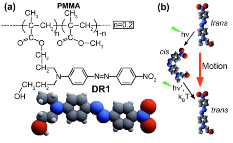
Fig. 1: (a) Molecular structure of PMMA-DR1. (b) Schematic of the trans-cis-trans isomerization cycle.
We applied this technique to the imaging of the optical intensity in the vicinity of chiral plasmonic structures [2]. As a canonical chiral structure, we studied two coupled gold nanorods, one rod being shifted with respect to the other. Changing the value of the shift s also changes the geometrical chirality, from an achiral structure at s=0 to chiral structures for non-zero values. A representative example of photochemical imaging on shifted gold nanorods is shown in Fig. 2. Here the nanostructures were illuminated using left-handed circular polarization (LCP), leading to the presence of a hot spot in the gaps between the nanorods. This hot spot is switched off under illumination with right-handed circular polarization. The images are compared with FDTD simulations of the electric field intensity, showing excellent agreement.
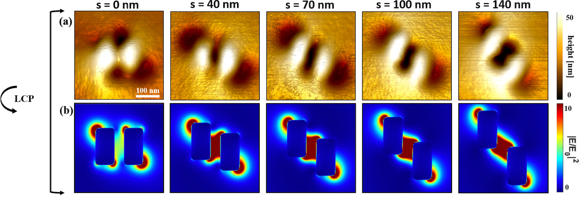
Fig. 2: (a) Photochemical imaging of laterally shifted gold nanorods with PMMA-DR1, for different values of the shift s. The structure is excited at 780 nm under LCP. (b) FDTD calculations of the electric field intensity. Adapted from [2].
Chirality in achiral nanostructures
Interestingly, chirality can also appear in geometrically achiral nanostructures [3]. The structure here is a metamolecule consisting of three gold nanodisks. The structure is obviously not chiral. However, under circularly-polarized excitation at a proper wavelength, interference effects between the eigenmodes of the structure yields to the electric field distributions that are mirror images of each other (see Fig. 3). We were able to imprint this "near-field chirality" in our photopolymer, turning an initially achiral structure into a chiral one.
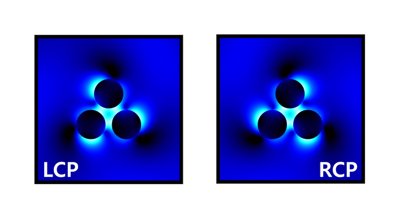
Fig 3: FDTD-calculated electric field intensity near three gold nanodisks (diameter D=140 nm, height 50 nm, gap distance 30 nm), illuminated at λ=770 nm, under RCP and LCP polarizations. Adapted from [3].
References
[1] Plain, J., Wiederrecht, G. P., Gray, S. K., Royer, P., & Bachelot, R. (2013). Multiscale Optical Imaging of Complex Fields Based on the Use of Azobenzene Nanomotors. The Journal of Physical Chemistry Letters, 4(13), 2124–2132[2] Aoudjit, T., Horrer, A., Kostcheev, S., Bachelot, R., Plain, J., & Gérard, D. (2023). Photochemical Imaging of Near‐Field and Dissymmetry Factor in Chiral Nanostructures. Advanced Optical Materials, 11(9), 2203015.
[3] Horrer, A., Zhang, Y., Gérard, D., Béal, J., Kociak, M., Plain, J., & Bachelot, R. (2019). Local optical chirality induced by near-field mode interference in achiral plasmonic metamolecules. Nano Letters, 20(1), 509-516.
mise à jour le 15 mars 2024