In the same section
Nanospectroscopy
Leading Scientist: Pr. Pierre-Michel Adam
Summary
This research topic aims to develop new spectroscopic techniques to characterize nanoparticles and their interaction with the environment. It includes extinction or scattering measurements of ensembles of nano-objects, as well as single nanoparticles. The development of specific techniques, like Fourier plane imaging or in-situ optical measurements during strain application, allows a deeper understanding of surface plasmon resonance phenomena or nano-antenna behavior and paves the way towards the development of highly sensitive sensors (bio-, chemico- or strain-sensors).The coupling of nano-systems with emitters (quantum dots or molecules) is also characterized via photoluminescence measurements by means of single photon counting system and lifetime resolved imaging. Finally, Surface Enhanced Raman Scattering detection allows us to characterize the presence of different molecules on surfaces, as well as the ability of nano-systems to enhance these drastically low spectroscopic signals.
Specific research topics & applications
Non-exhaustive list
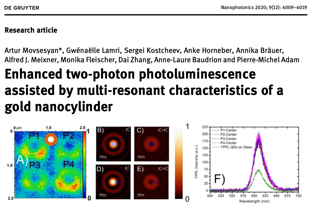
- Non-linear nano-optics (leaders: A-L Baudrion & P-M Adam)
- Biosensors (leader: E. Ionescu)
- Strain sensors (leader: T. Maurer)
- Smart lighting devices (leader: A-L. Baudrion)
- Surface enhanced Raman scattering (leaders: S. Jradi and P.-M. Adam)
Highlights

Equipments:
Single nanoparticle scattering measurements:
Based on a Zeiss Axio Imager Z2 microscope, this system allows us to detect the scattering signal of single nanoparticles. Illuminated either by a Halogen or by a Xenon lamp through a darkfield objective, the scattered signal is collected and sent to a spectrometer via an optical fiber, whose core acts as a confocal filter, resulting in a very high signal to noise ratio. This system can be used either in reflection mode or in transmission mode, where the illumination is performed by a darkfield condenser in oil, leading to a Total Internal Reflection illumination.
Angle resolved extinction measurements:
This home-built system lies on the use of objectives with long working distances. Indeed, these illumination and collection objectives are far enough from the sample to place the sample holder on a goniometric stage. This allow to tilt the sample in the optical axis and to record extinction spectra under oblique incidence. The illumination is performed via a Xenon lamp and the detection by a fiber-coupled spectrometer (QE6500, Ocean Optics).
Scattering diagram imaging:
A filtered supercontinuum laser is used to illuminate nanoparticles at the desired wavelength. The scattered light emitted by the sample is collected by an oil immersion objective, whose back focal plane is imaged on a CCD Camera. Some intermediate lenses are added in order to add a beam blocker in an intermediate back focal plane image. This allow to filter the incident laser beam and to collect only the scattered light in a given range of emitted angle (typically from 39 to 73°).
Micro-Raman spectrometer:
The system makes possible it to perform Raman scattering measurements with sub-micrometric lateral and vertical resolutions. This commercial set-up is composed of an optical microscope coupled with a highly sensitive spectrometer. It can be operated both in a local spectroscopic mode and in a scanning imaging mode. The system is equipped with five different lasers, whose wavelength ranges from near-UV (325nm) up to near-IR (785nm), with three visible wavelengths in between (405nm, 532nm, 633nm). Control of linear polarizations is available for all wavelengths in both illumination and collection paths. Circular polarizations and low-frequency Raman signal detection is also possible. The Raman microscope is equipped with a heating/cooling cell.
Fluorescence Lifetime Imaging Microscope (FLIM):
The FLIM system is based on a Zeiss Axio Observer inverted microscope, mounted with the Becker & Hickl DSC 120 confocal FLIM. Three picosecond laser lines (405, 445 and 473 nm), as well as the SuperK Extreme EXR20 supercontinuum laser (NKT Photonics) can be used to excite fluorescent systems. The laser is scanned on the sample and the fluorescent signal is detected at each point. The Time Correlated Single Photon Counting (TCSPC) module is responsible for tagging each photon with its travel time since the pulse start and the fluorescence lifetime map is calculated via the SPCImage software. This system is very useful to analyse the influence of metallic nanoparticles on fluorescent entities.
Confocal Microscope:
The Zeiss LSM 780 confocal microscope is mounted with six continuous laser lines (405, 458, 488, 514, 561 and 633 nm). Two PMTs and a 32-channels-GaAsPs detectors are mounted in the spectral module, allowing to record simultaneous images at different emission wavelengths. Finally, the Z-stack module allow to record 3D-images of the fluorescent signal.Date of update 27 janvier 2022
Contact
Collaborations
- Monika Fleischer's group - Eberhard Karl Universität Tübingen , Germany
- Dai Zhang's group -Eberhard Karl Universität Tübingen , Germany
- Physics Department - Politecnico di Milano , Italy
- Institut Lumière Matière - Lyon, France
- ITODYS - Université Paris Diderot , France


